The Eye and How It Works
The first step in understanding glaucoma is to know a few basic facts about the eye and how it works. Once you have this information, you will be better able to discuss your condition and treatment with your eye doctor. Working together, you and your doctor will be able to act as a team to protect your vision.
Think of Your Eye as a Camera
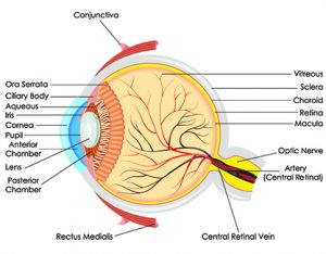
 The eye is like a camera. It has a lens which focuses light, just like the lens of a camera. In the eye the focused image is formed on the retina, in the back of the eye. The image information (color, shape and movement) is then sent to the brain via the optic nerve, which connects the eye to the brain. This is very similar to a digital camera, which can be connected to your computer via a computer cable, allowing the images to be transferred to your computer. In glaucoma, the lens and retina function normally, but the optic nerve is damaged and images cannot be transmitted to the brain.
Key Parts of the Visible Eye
Let’s look at the eye more closely. The sclera is the white outer surface of the eye, a thin, yet tough, protective outer shell, which is covered by the conjunctiva (white-colored outer skin of the eye that contains some blood vessels). At the center front of the eye is the cornea. It is a clear dome-like structure that covers the iris and pupil. Light rays enter the eye through the cornea.
The pigmented portion of the eye is called the iris. It is responsible for eye color. It also controls the size of the pupil, the dark-colored area in the center of the iris. Together, the iris and pupil act like the aperture of a camera, depending on the amount of light your eye senses. When there is a great deal of light, as outdoors on a sunny day, the iris constricts the pupil, making it smaller and limiting the amount of light which passes through the pupil to the retina. When there is little or no light, the iris dilates the pupil, widening it so that more light can enter the eye.
The lens, located immediately behind the iris, adjusts its shape and thickness to focus the light rays onto the retina. (Often, as we get older, the lens gets discolored or hazy, and it is then called a cataract. A cataract can affect the ability of the lens to focus.) The retina, lining the back of the eye, then delivers the image as nerve impulses via the optic nerve to the brain, which processes these signals into a visual image.
The space in the eye that is behind the cornea and in front of the iris is called the anterior chamber. It is filled with a water-like fluid called the aqueous humor, which nourishes the cornea and the lens, providing oxygen and vital nutrients. The aqueous humor also provides the necessary pressure to help maintain the shape of the eye. We call this intraocular pressure or IOP. As you will read, maintaining the right amount of pressure within the eye is very important to protecting your vision. Measuring the IOP is one of the ways your eye doctor tests for glaucoma.
